Abstract
Introduction: Bone marrow biopsy is a staple procedure that hematology/oncology fellows must master during their 3-year fellowship. It is crucial for trainees to be able to practice their skills in a safe manner prior to performing the procedure with supervision and/or independently. Knowing the anatomy of how to perform this procedure is of utmost importance in order to avoid adverse outcomes such as severe retroperitoneal hemorrhage.1,3 Many fellowships do not have a standardized way to teach bone marrow biopsies and has traditionally been "see one, do one, and teach one," while other programs incorporate mannequins, cadaver, or 3D printed models for fellows to practice on.2 Case Western Reserve University is one of the 1st medical schools in the country to incorporate HoloAnatomy mixed-reality learning for anatomy training for their 1st year medical students. A subsequent randomized study published in JAMA confirmed that medical students did prefer mixed-reality for learning anatomy and also subsequently scored higher on their testing as well.4 We are the 1st hematology/oncology program in the country to incorporate mixed-reality learning for fellow training of bone marrow biopsies.
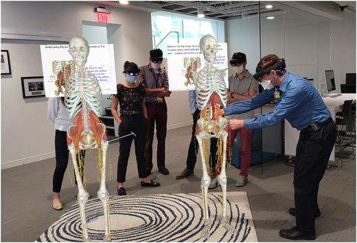
Methods: This was a pilot study conducted at the University Hospitals/Case Western Reserve University hematology/oncology fellowship program involving their 1st year fellow class made up of 5 individuals. Participants were asked to fill out a pre-test regarding their confidence in performing the procedure, observation of procedure in the past, optimal landmark(s), chronological order of correct steps of performing procedure, and whether they thought a combination of mixed-reality learning and mannequin training would help them learn the procedure better. They then were introduced to HoloAnatomy and had a 30 minute session regarding the anatomy of the pelvis from a certified anatomy instructor at CWRU- including going over the regional nerves, blood vessels, muscles and were able to visualize where their instrument would be directed for the biopsy. This was followed by a short 15 minute session from our fellowship faculty who introduced them to the bone marrow biopsy kits available at our institution and then observed two experts perform the procedure on a mannequin. Each individual was then allowed to perform the procedure under supervision on the mannequin. Participants were then asked to complete the post-test regarding their experience at the conclusion of the workshop.
Results: A total of 5 individuals participated in the pilot study (males n = 3, females n =2). Four of the 5 individuals came directly from graduating internal medicine residency and 1 individual had been a hospitalist for 3 years. The pre-workshop results showed that the average confidence in performing a bone marrow biopsy was 1.4 (graded from 1-10) and post-test revealed this went up to an average of 5 (graded 1-10). Prior to the workshop, the average number of observed bone marrow biopsies was 5 (range of 2-10). 100% of participants in both the pre and post settings knew the optimal landmark was the posterior superior iliac spine. The pre-test showed 4/5 participants were able to identify what steps not do prior to performing a bone marrow biopsy. The pre-test average score was 54% (range 43%-71%) for correctly identifying the correct chronological order of steps to perform a bone marrow biopsy, while post-test revealed the average increased to 80% (range 43%-100%). 100% of participants also felt that the combination of virtual reality and mannequin training led them to becoming more comfortable with the procedure.
Conclusions:
HoloAnatomy/mixed reality anatomy training is a novel way of introducing anatomy to not only medical students, but also later post-graduate medical trainees in performing procedures, such as a bone marrow biopsy. We are the 1st hematology/oncology fellowship program to incorporate this into our curriculum and use a combination of mixed reality anatomy and mannequin training to prepare our fellows to perform this procedure. We have objective evidence in the medical student world and now in the post-graduate training world that the combination of mannequin and mixed reality anatomy training can further improve and augment our medical trainee's learning experiences in performing procedures such as a bone marrow biopsy. We hope that this will become more of a standard moving forward in all post-graduate medical training.
Disclosures
No relevant conflicts of interest to declare.
Author notes
Asterisk with author names denotes non-ASH members.


